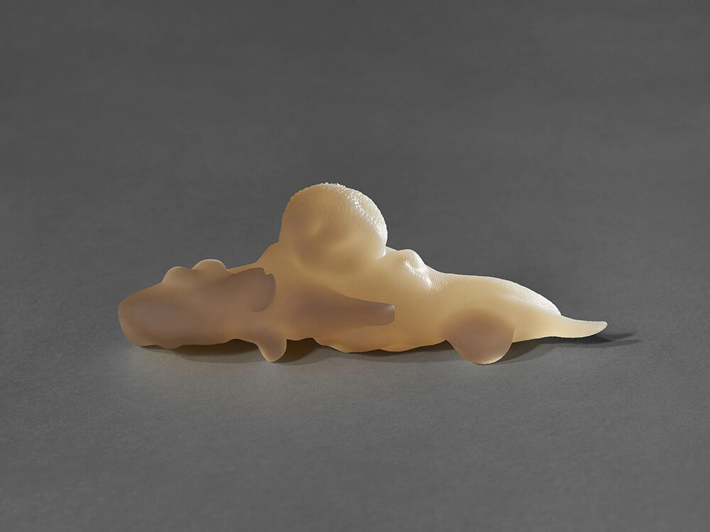
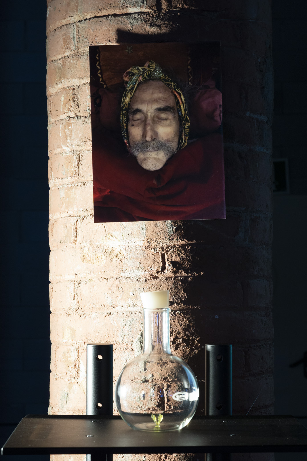
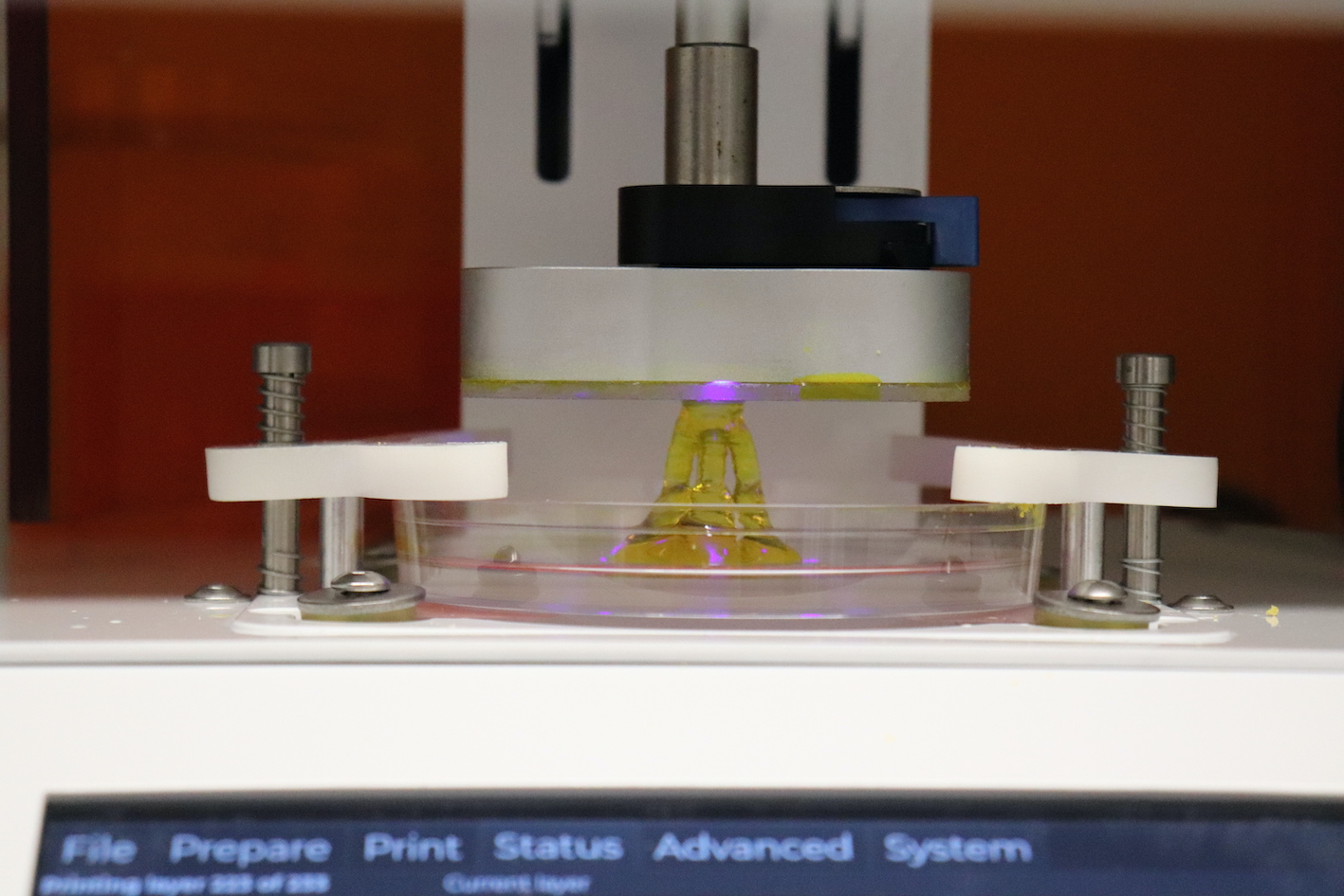
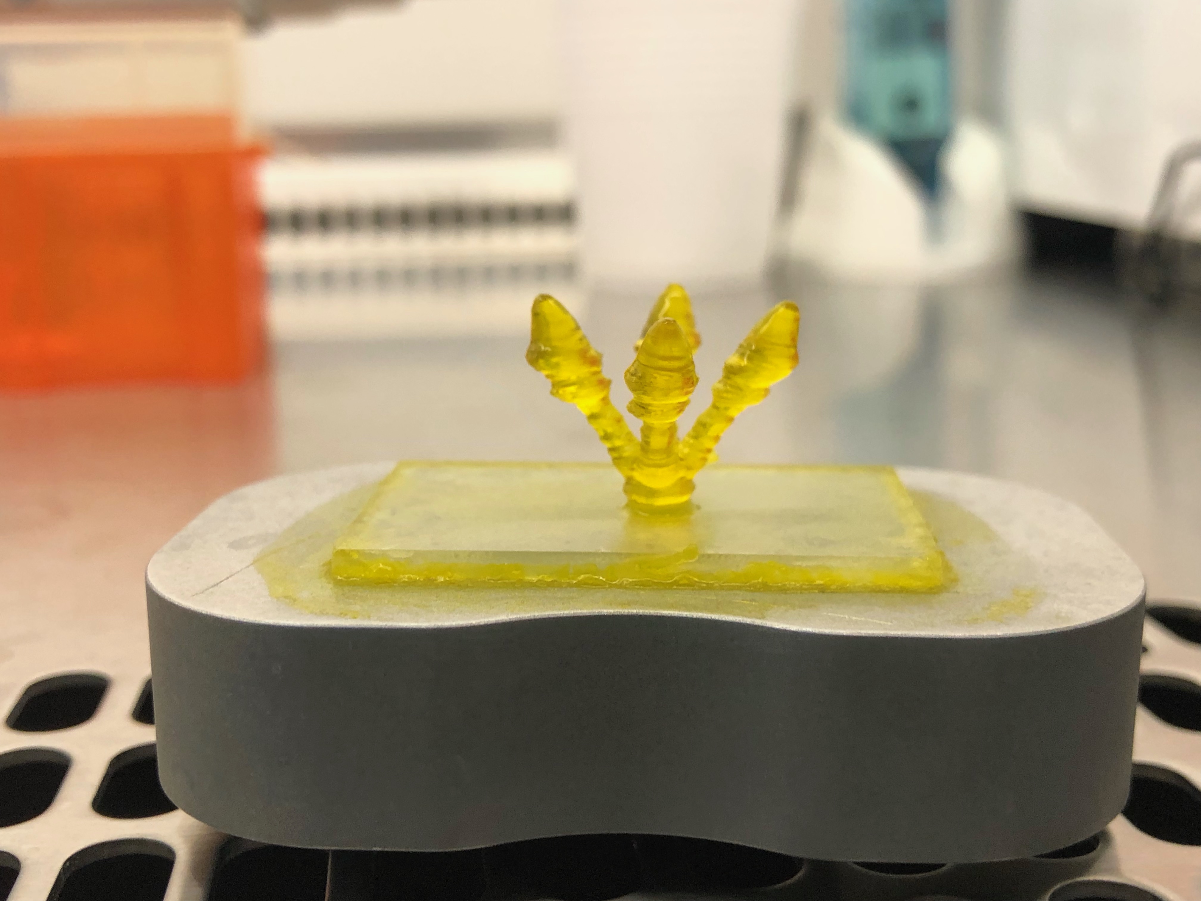
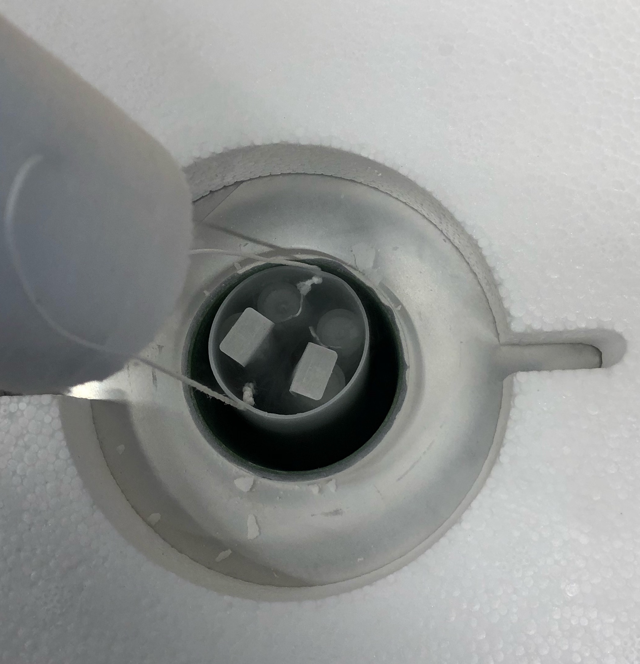
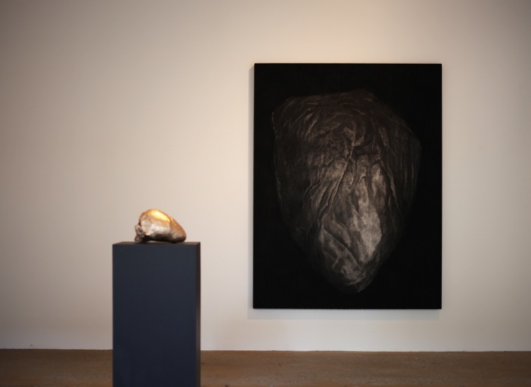
Documentation from Last Breaths installation at ISEA 2022
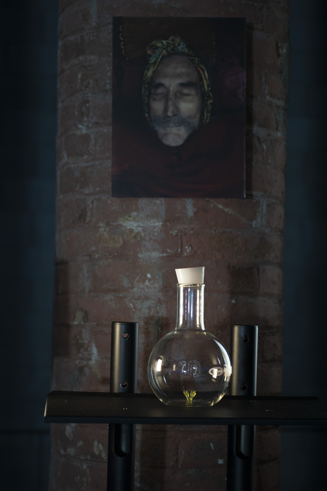
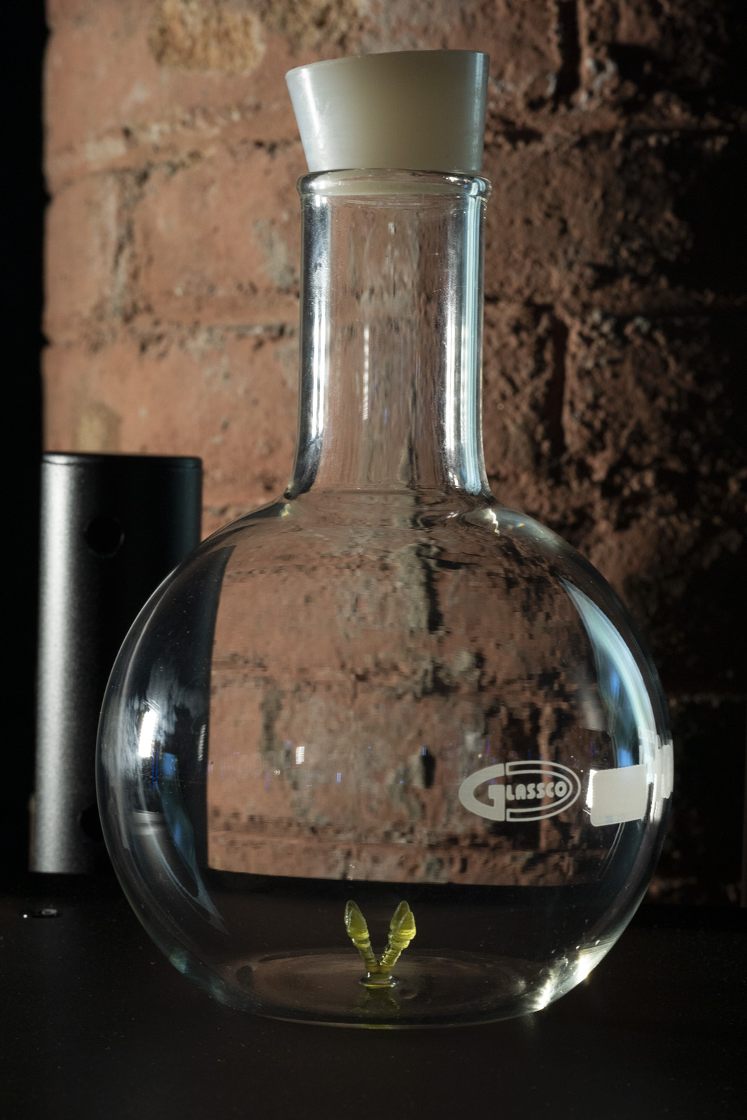
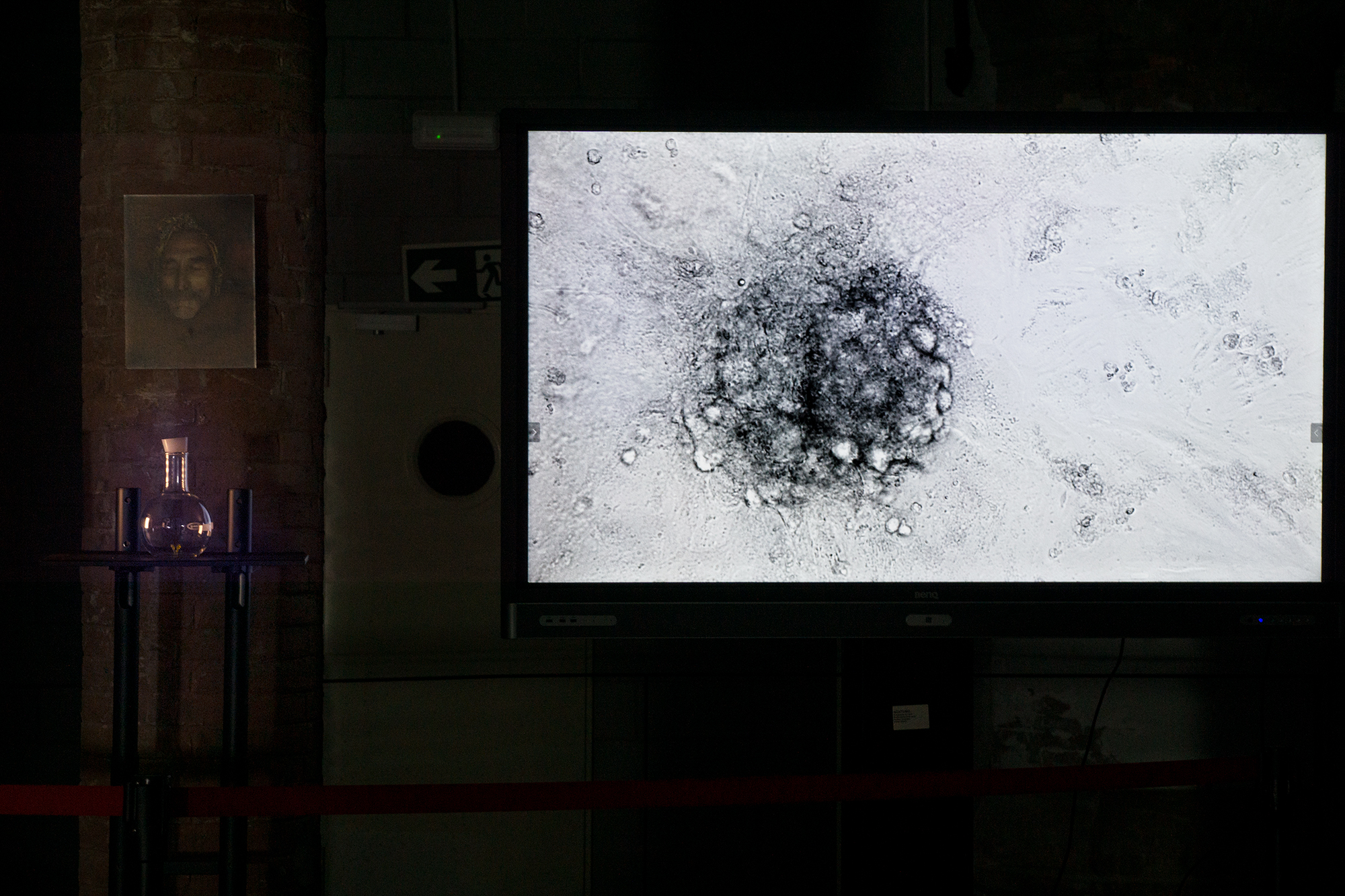
Last Breaths installation with bioprint, photo of George Schwarz, video including beating cardiac spheroid microscopy by Clara Liu Chung Ming.
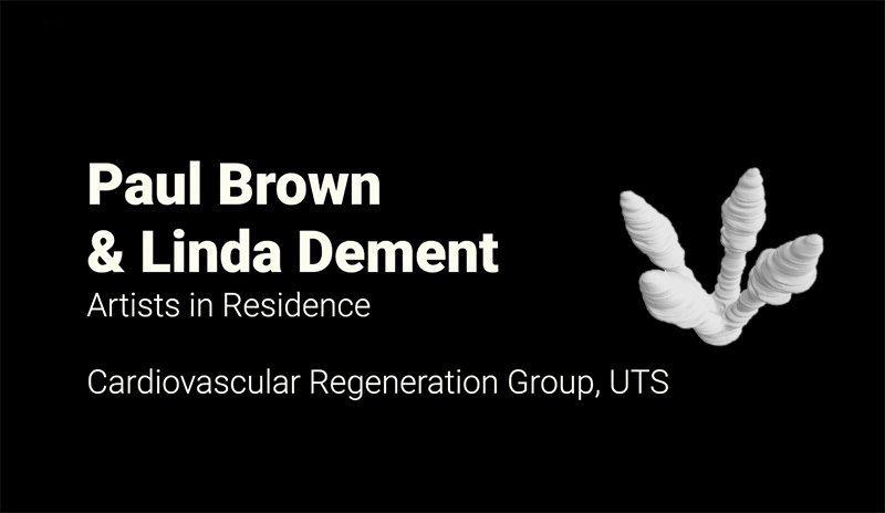
Paul Brown + Linda Dement – Heart Project
ANAT Synapse Residency 2021
Sunday, 3 July 2022
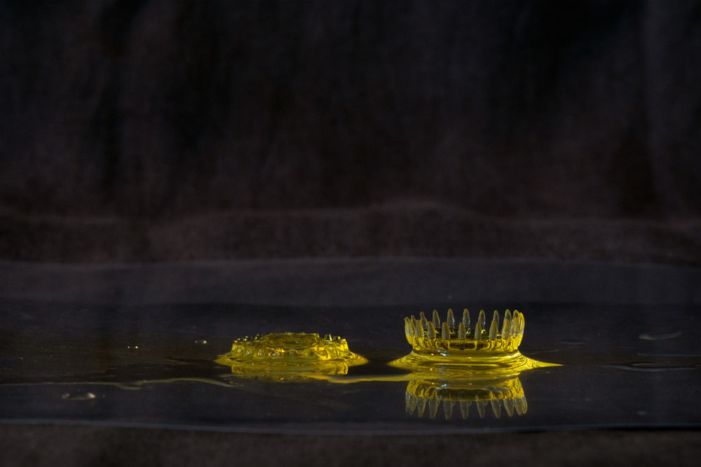
Documentation from Last Breaths installation at ISEA 2022
Last Breaths installation with bioprint, photo of George Schwarz, video including beating cardiac spheroid microscopy by Clara Liu Chung Ming.
Monday, 13 June 2022
Our exhibit for ISEA 2022 opened on Friday night, and will run until 30 June. The venue is the Recinte Modernista de Sant Pau, one of several housing this year’s ISEA exhibitions. Meanwhile the ISEA Symposium is on, and Linda and Carmine are attending the sessions.
For more about ‘Last Breaths’ go to: http://ISEA Possibles exhibition
Last Breaths
Linda Dement, Paul Brown, Carmine Gentile 2022
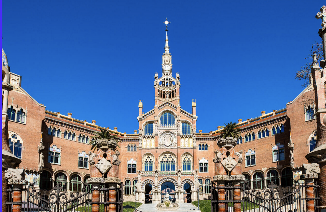
Recinte Modernista de Sant Pau, Barcelona – formerly a hospital. Venue of Possibles exhibition, ISEA 2022
Friday, 3 June 2022
Exiting times for our project–Linda and Carmine fly out this weekend to install our sculpture ‘Last Breaths’ for ISEA 2022 in Barcelona. The ISEA exhibition runs 9-30 June in multiple venues.
We’re privileged to be selected by the jury for installation at the historic Sant Pau hospital in central Barcelona. ISEA (International Symposium on Electronic Arts) is a prestigious annual symposium and exhibition, dedicated to the intersection between art, science, technology, design and engineering.
See more about ISEA 2022 on facebook: https://www.facebook.com/ISEA2022Barcelona/
Find the web page for ‘Last Breaths’ among the exhibits: https://isea2022.isea-international.org/event/sant-pau/
Linda: “Through this project, human cardiac failure, expelled as breath and recorded as sound, is transfigured to 3D form as terra-firma for growth of human yet inhuman life. The work traverses terrains of life and death, bodies and technologies, human and inhuman, the strained edges of wounded hearts and the stretching extremities of bio-technical possibility.”
We’ll report from Barcelona over the next two weeks.
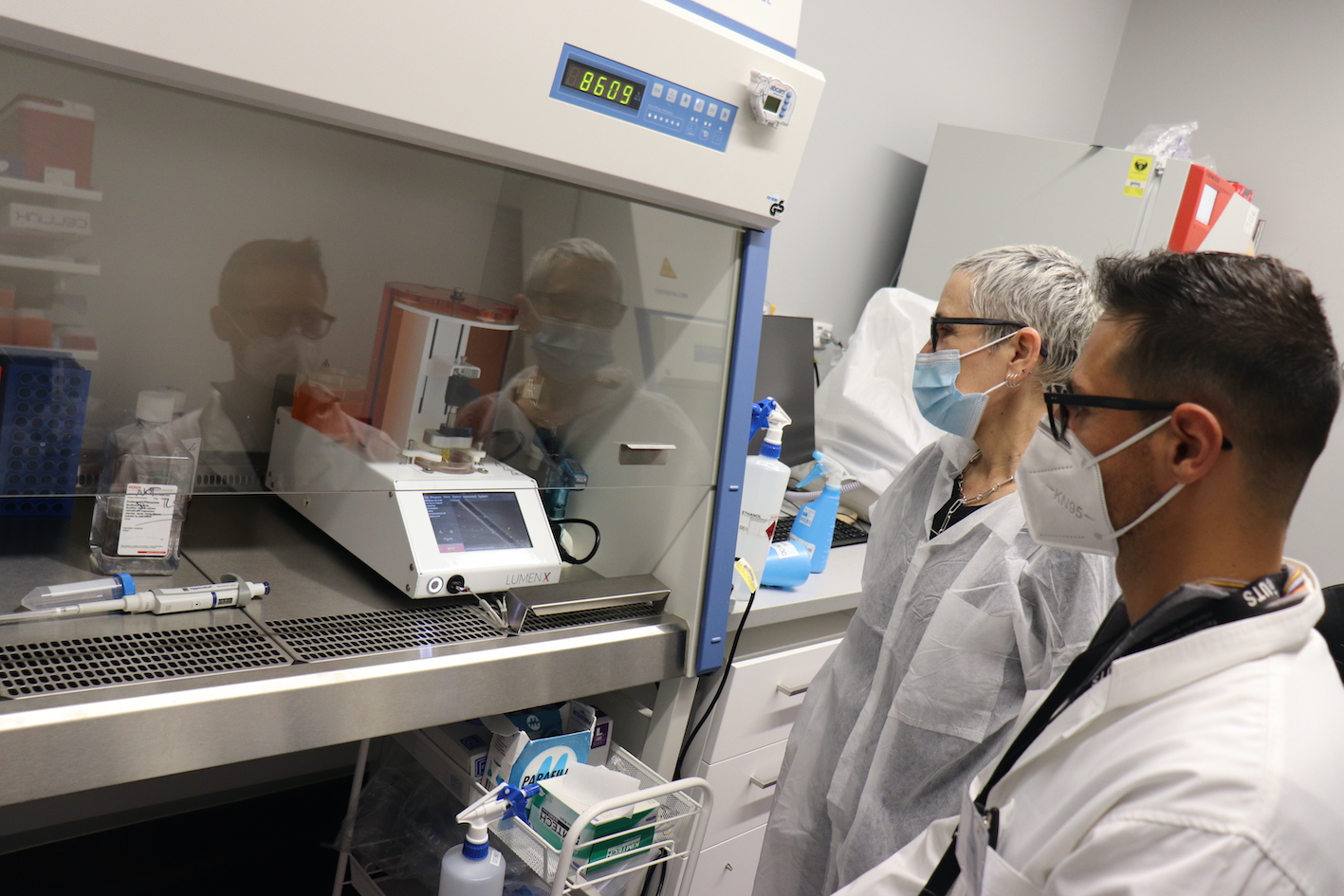
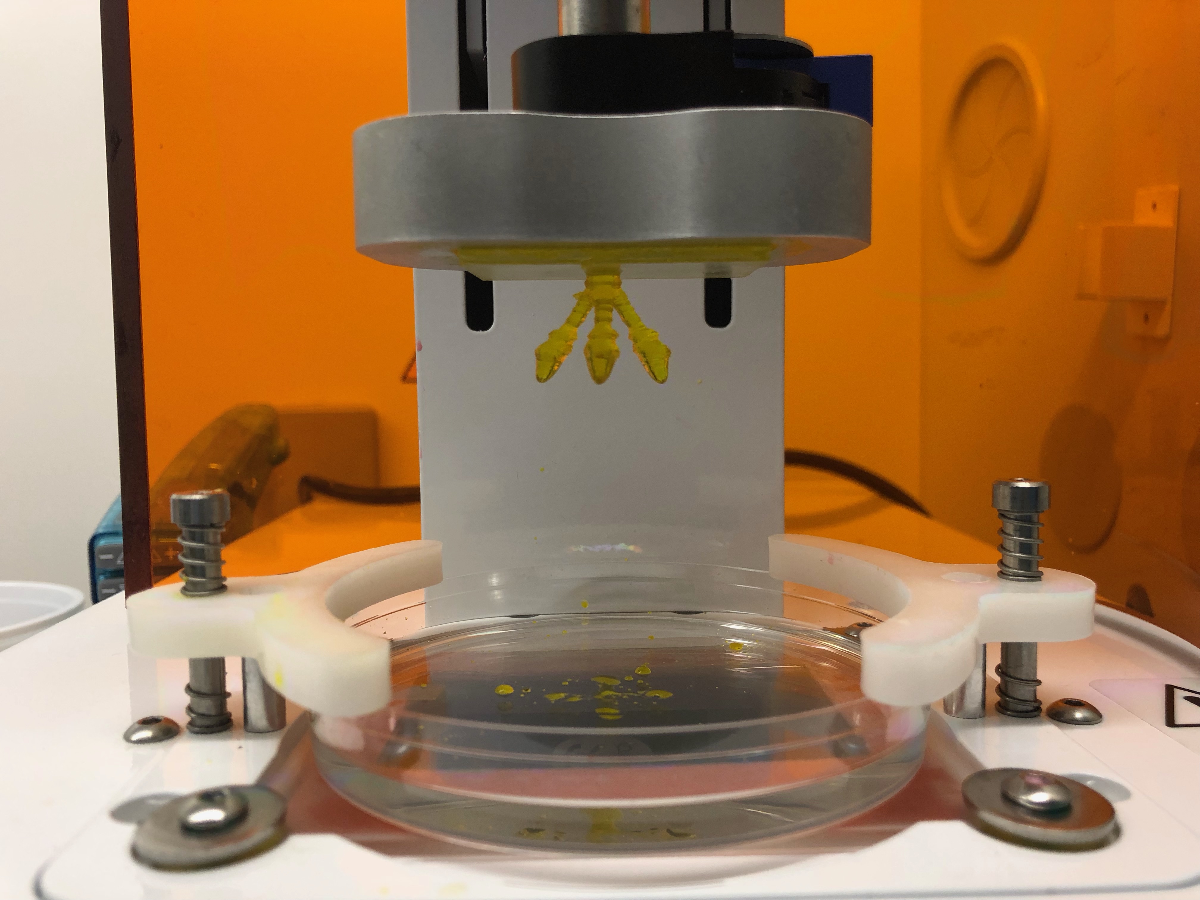
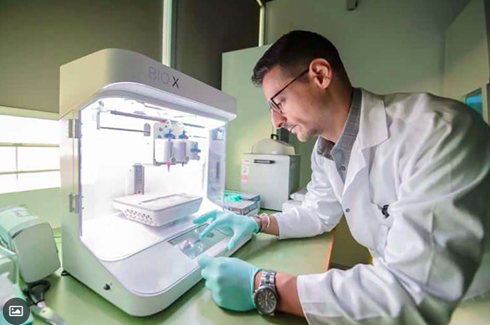
Bioporinting with the LumenX in the UTS lab, Carmine Gentile and Linda Dement
Printing underway in LumenX bioprinter
Bioprint completed in the LumenX bioprinter
Wednesday, 30 March 2022
We have completed the work on the Heart Project for 2021, although our collaboration with the UTS Cardiovascular Regeneration Group is continuing and we’ll continue to update this blog.
At this point we want to again acknowledge the support of Dr Carmine Gentile and his laboratory team at UTS, as well as the generous financial support from our funding organisations, ANAT via its Synapse program, and Create NSW.
Proudly funded by the NSW Government in association with the Australian Network for Art and Technology
![]()
![]() Cardiovascular Regeneration Group
Cardiovascular Regeneration Group
![]()
Thursday, 27 January 2022
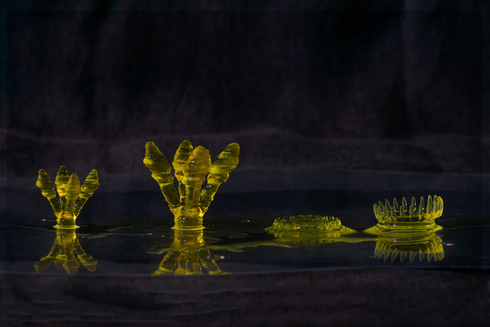
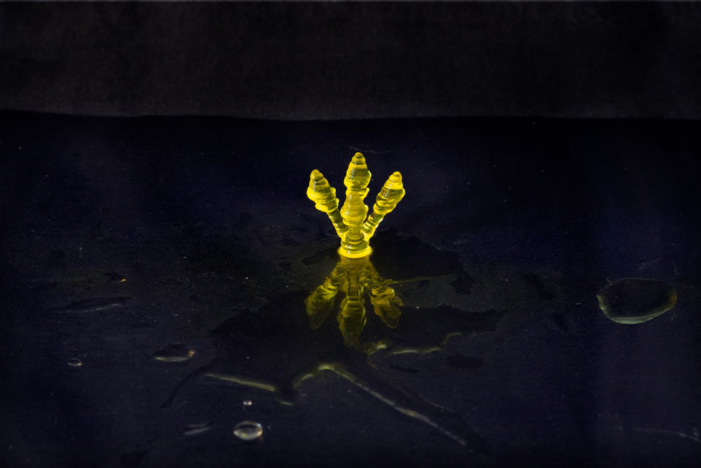
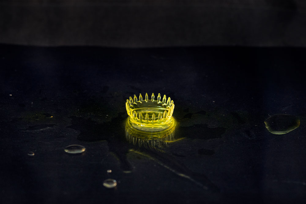
Towards the end of 2021 we were able to go with Dr. Carmine Gentile into the lab a few times, where he printed the different forms and at different sizes, in hydrogel.
These experiments will enable us to work out which form, at what size and what printer settings to print with live cells.
We later spent a day photographing the hydrogel 3D prints:
Thursday, 9 December 2021
Pleased to be able to undertake more experiments in the lab, after lockdown restrictions eased. We aim to produce a series of sculptures, to contain live cells embedded in hydrogel (alginate gelatin). We are documenting the lab work, aiming to produce short time-lapse films of the process. Here is a trial run…
Thursday, 18 November 2021
Our next steps might be to print one or more of these forms with cells. We are interested in finding out:
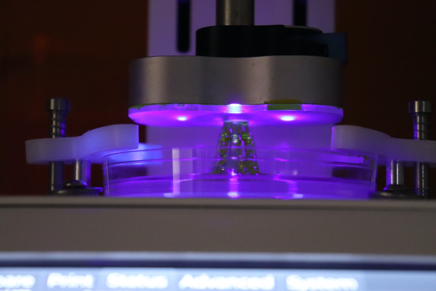
We’d like to video the next round of bioprinting, maybe with time-lapse, to see the form built layer by layer in the LumenX printer. Not much is visible, especially at first, except darkness and tiny purple lights exposing the gel. We’ll have to find a way to take a tripod and camera into the lab and set it up in the darkened room.
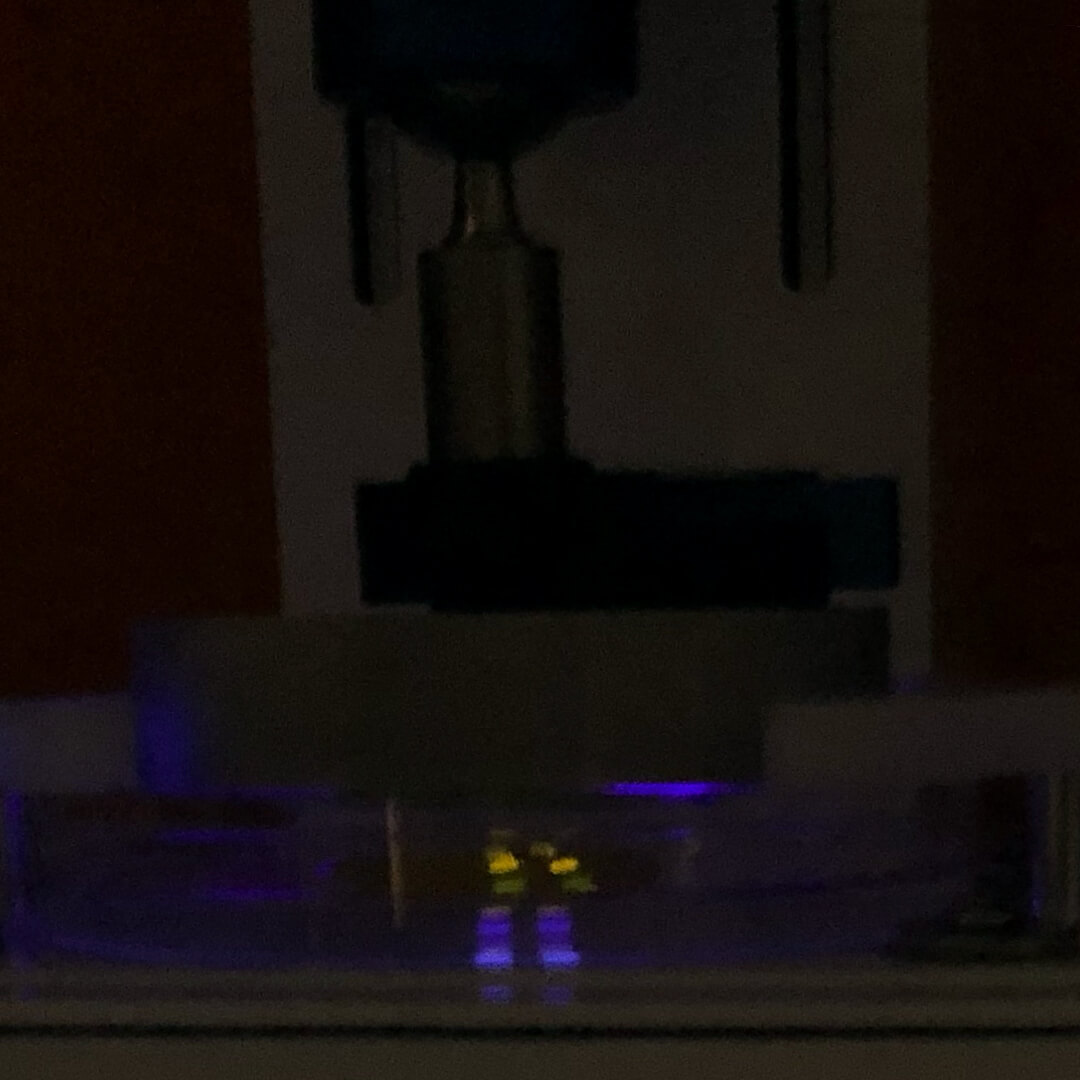
the LumenX underway
The procedure is fascinating.
From the LumenX site:
The Lumen X+ divides 3D models into stacks of horizontal layers in the form of black and white image files. Using an industrial-grade visible-light projector, each image is projected onto a droplet of liquid PhotoInk™ on a polydimethylsiloxane (PDMS) vat. The illuminated regions react and solidify, then the build platform moves the cured layer up and out of the way, so that more liquid PhotoInk can be cured with the next image.
So the form appears to be drawn upwards and out of a blob of liquid on the plate, the purple lights conjuring substance out of fluid.
Thursday, 4 November 2021
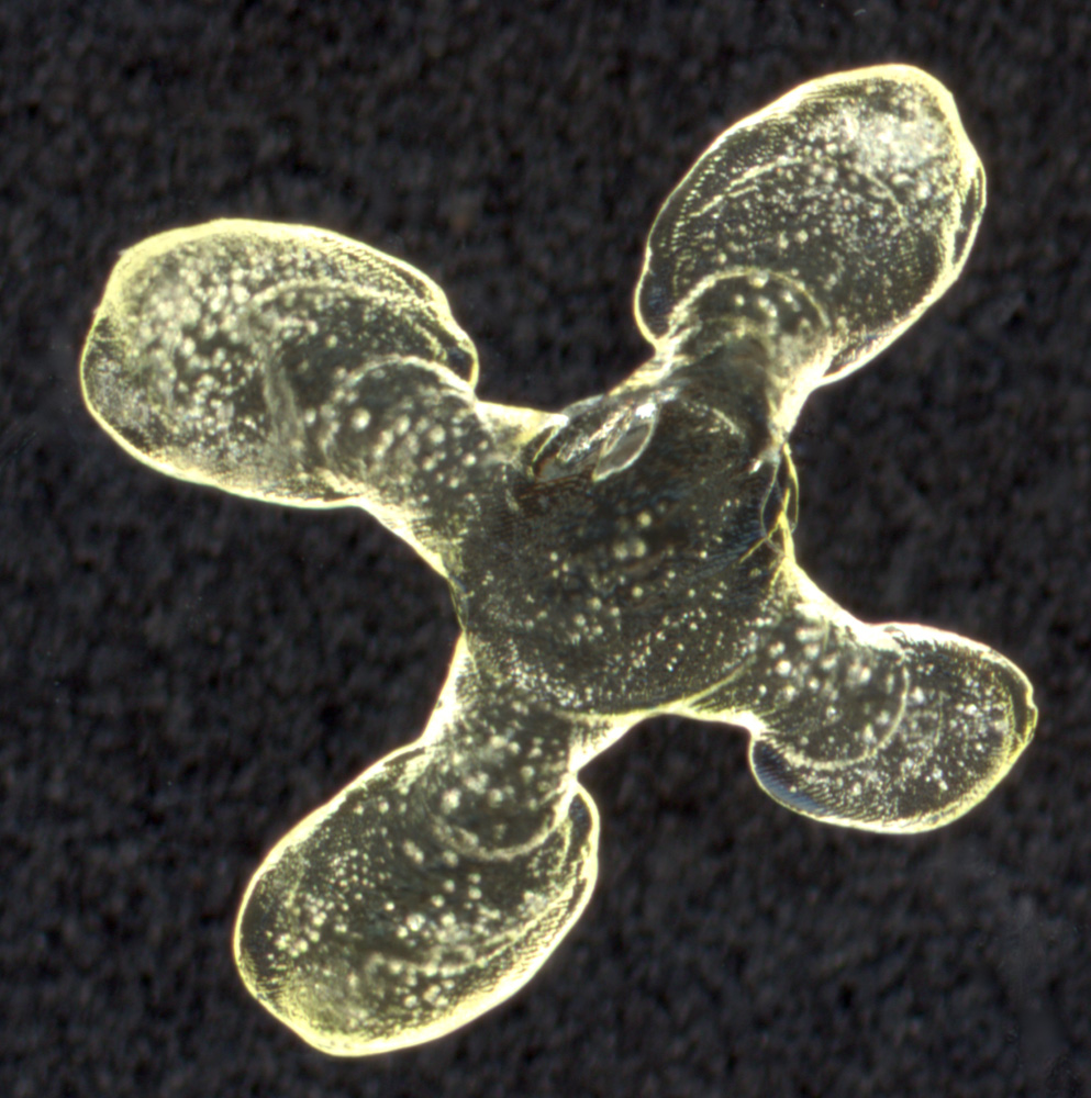
Extra images from our recent trials of mini-sculptures.
Photographs by Dr Carmine Gentile and team at the Cardiovascular Regeneration Laboratory
Friday, 22 October 2021
Carmine Gentile set up the LumenX bioprinter and did some trial prints of our shape.
The LumenX uses crossbeams of light to harden a light sensitive gel. The printer starts at the bottom and does a thin layer (100 microns) then lifts up and does the next layer and so on. The non-hardened gel drops away and the form is left. He experimented with the length of time that the light was on and the intensity of the light to get an exposure that would harden the gel adequately – very much like printing a photo in the darkroom.
I used to love working in the darkroom. I’d do endless test strips with sections exposed for different durations. In the colour lab I liked it when I could have 2 enlargers going at the same time. Expose #1 and put it through the machine, expose the next. By the time I’d done exposure #2, test strip #1 was ready to look at. I could lose days in there and rarely see sunlight. That’s the one thing I miss about working digitally.
In photography, I would have expected that 1 image from every 2 rolls of film would be successful – an image I would want to use. I was mostly working with an old medium format Mamiya which had an anomaly that I liked, it did 11 images per roll though it’s supposed to do 12. That’s a 1 in 22 ratio. Some weeks or even months on end, when the stars aligned in a fortunate manner, I would get 1 in 11.
That 1 in 22 or 1 in 11 is not luck. Quantity is necessary to experiment, develop and refine to find quality. Failures are crucial.
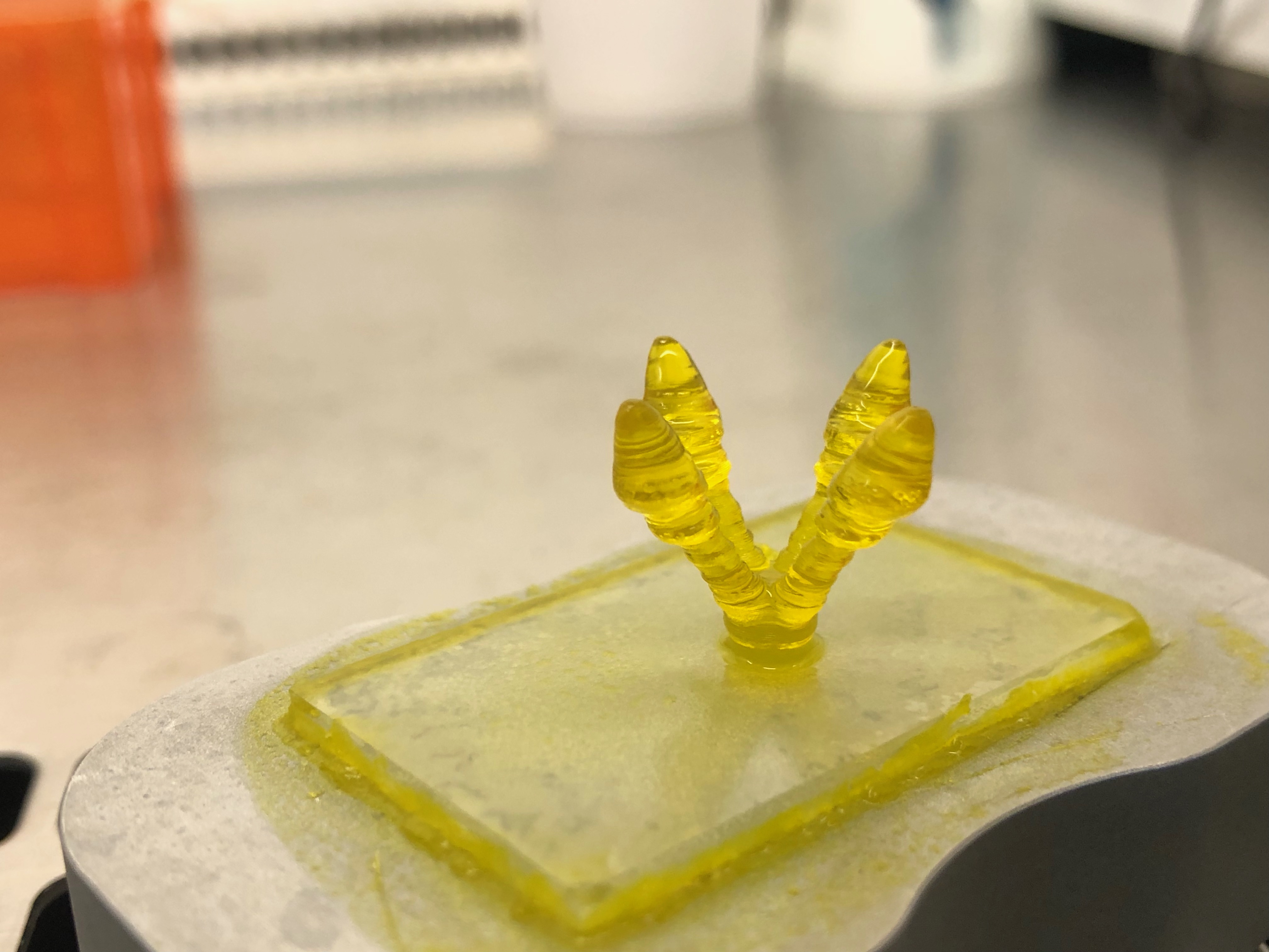
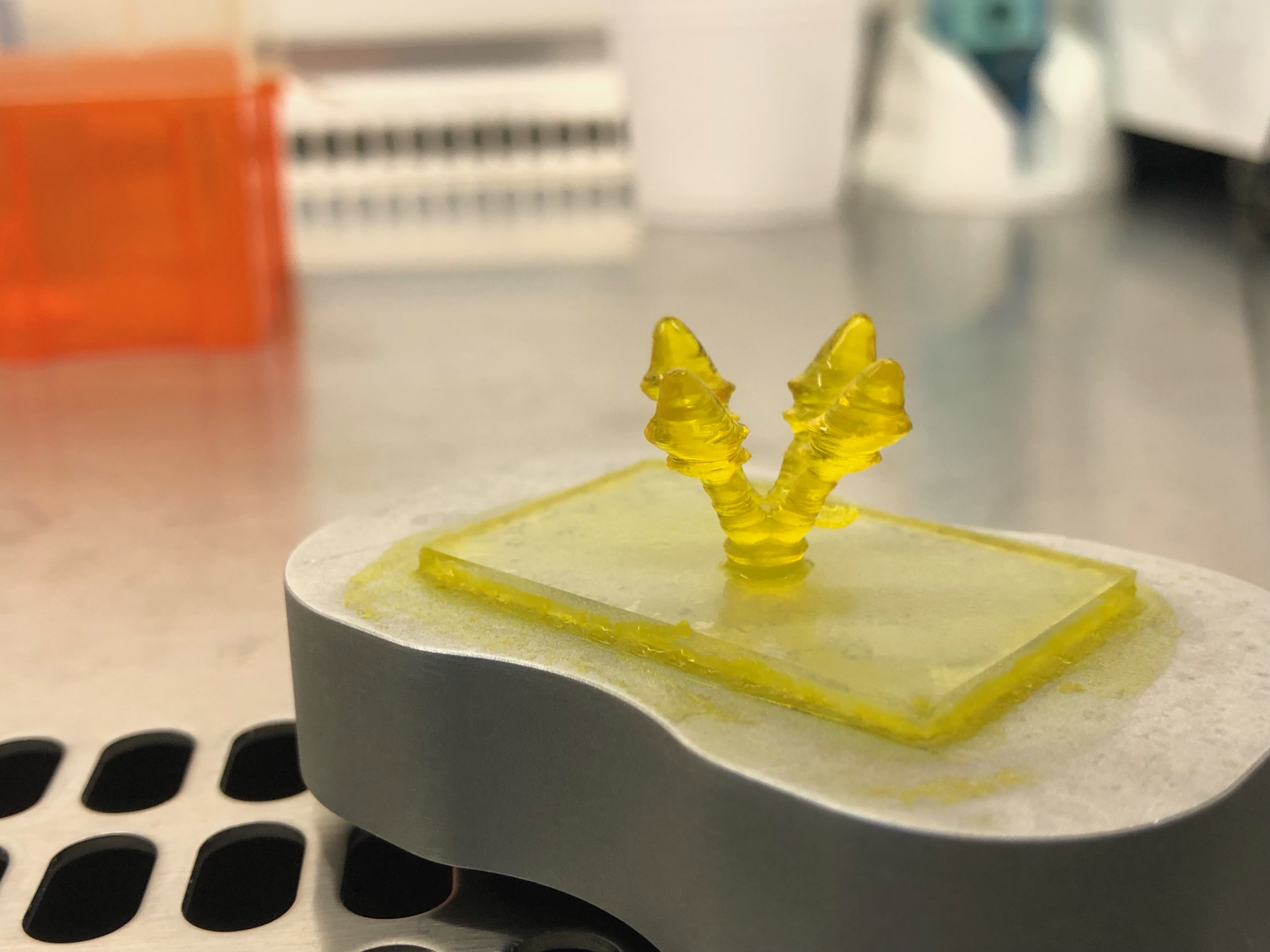
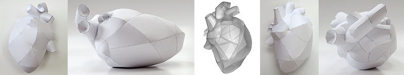
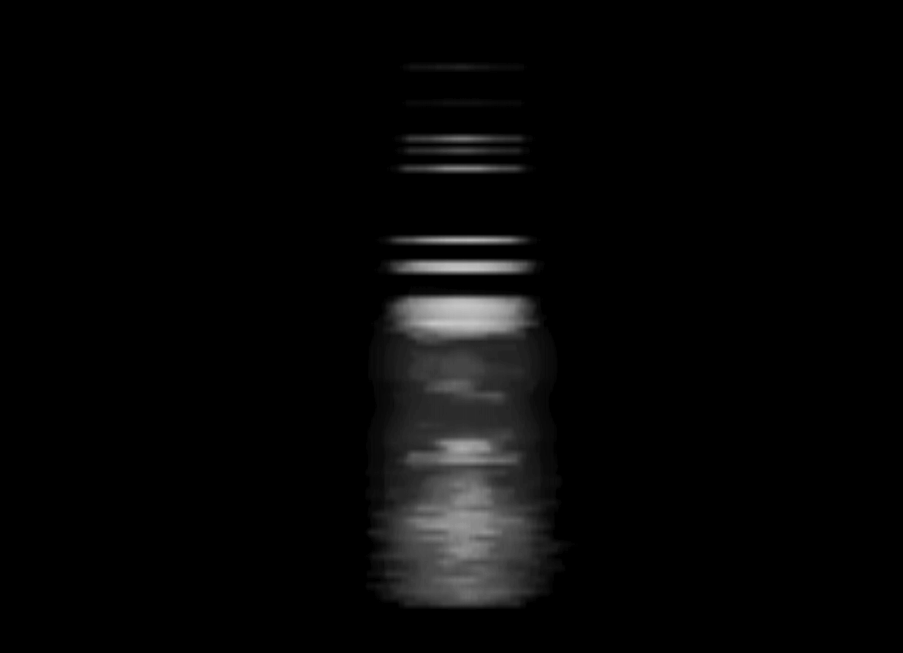
The first two LumenX printer runs left a flat little gum drop of a thing:
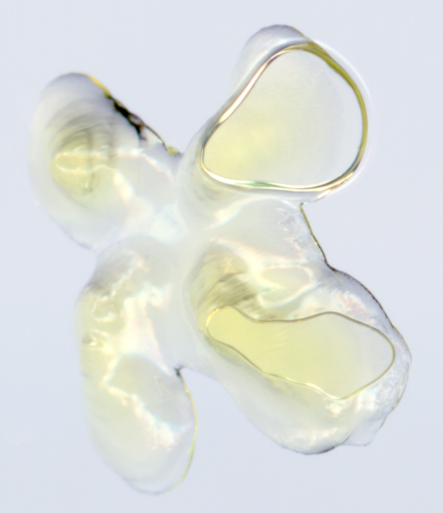
It had collapsed in on itself with each new layer as it wasn’t hard enough. The third print was a success. This glorious little object is 1cm high and golden.
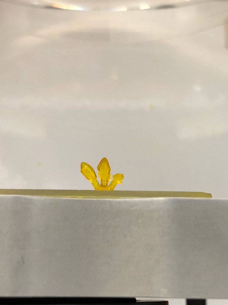
photo: Carmine Gentile
Friday, 15 October 2021
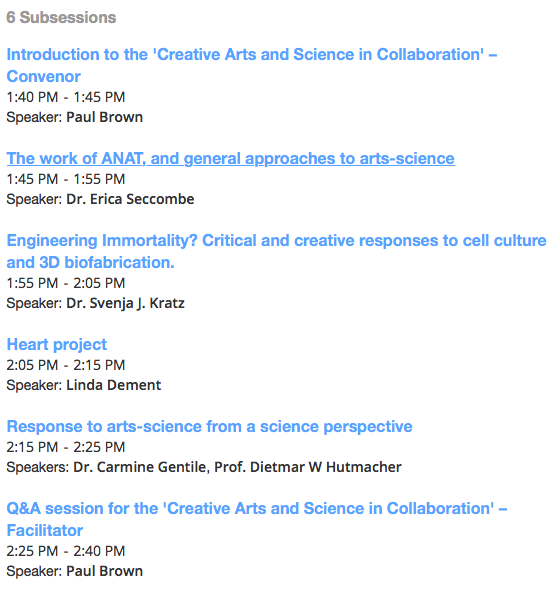
The 3rd Australian Bioprinting Workshop took place on 4-5 October. A great success! 550 registrations and 50 speakers from around the world.
Much positive feedback for our presentations about arts-science and artists’ contributions to bioprinting – a great way to bring together the current artists for the Synapse residencies for 2021.
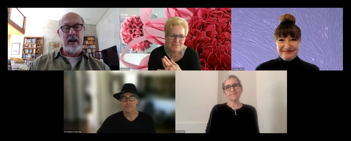
Paul chaired the session, and ANAT Board member Erica Seccombe started us off with an overview of arts-science connections and the reason why ‘A’ for Arts is so important to include in STEM, to make STEAM!
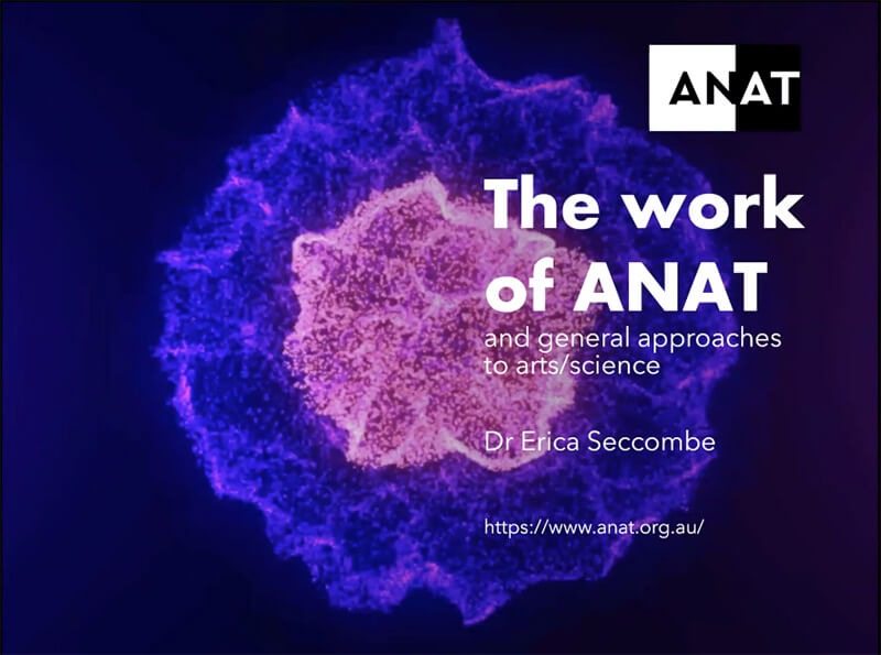
Erica Seccombe: The work of ANAT and general approaches to arts-science
Svenja Kratz gave her talk on her current Synapse project and Dieter Hutmacher and Carmine Gentile responded, as the scientist collaborators for Svenja and for our own project.
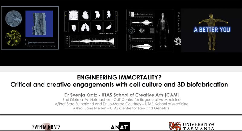
Svenja Kratz: Engineering Immortality? Critical and Creative responses to 3d biofabrication
Linda presented on our own project and our progress to date in the terrain between life and non-living, form and immateriality.
Paul Brown and Linda Dement: The Heart project, in residence with Dr. Carmine Gentile’s Cardiovascular Regeneration Group
For the whole 3rd Australian Bioprinting Workshop, presentation recordings are now available on-demand and can be accessed directly through the Australian Bioprinting Workshop site.
And some other great news!! As COVID restrictions lift, we will soon be able to visit the UTS lab, and move on with producing 3D sculptures. Onwards and upwards!
Friday, 24 September 2021
We are contributing to an amazing two-day conference/workshop coming up on 4-5 October. The 3rd Australian Bioprinting Workshop for Tissue Engineering and Regenerative Medicine is a FREE on-line webinar hosted by Carmine Gentile from the UTS Cardiovascular Regeneration laboratory, and will include a session on the role of the arts.
With us will be Svenja Kratz, also a Synapse Resident this year, and Erica Seccombe from ANAT’s board, plus commentary from scientists Carmine Gentile and Dietmar Hutmacher.
Why not join us! On-line access for the 3RD Australian Bioprinting Workshop
Here’s a glimpse at the Art-Science workshop session, which is on Tuesday 5 October starting at 1.40pm.
Thursday, 23 September 2021
It started Monday 13 September with international despatch from Wisconsin USA of 8 vials of human heart cells – 40 million cells in all. By Wednesday they’d reached the FedEx depot at Matraville in Sydney’s south east. But they were stuck in Customs. On Friday nothing had changed – except most likely the dry ice and liquid nitrogen controlling temperature inside the parcels would be degraded, and the cells can’t survive if they overheat.
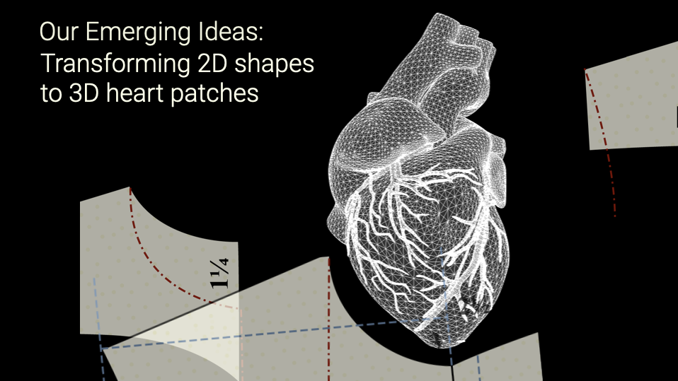
Across that week, we also had key discussions with collaborating paper artist Horst Kiechle, and with PhD student Niina Mathews – continuing our development of ideas about the flexibility, the dimensions, the shapes, of heart patches. Importantly, we progressed our thinking about how to present creative works, and had very helpful discussion with the UTS Gallery.
By the following Monday afternoon, this week, the cells – which we intend to include as the living element in mini bio-printed sculptures – were still stuck at the depot, though cleared by Customs. What next? We got news that our two packages had been separated, that one was on its way to another depot in a semi trailer.
Tuesday morning, masked, double-vaccinated, and ‘travelling for work’, Carmine and Paul hit the road to liberate the cells from the depot – well it seemed that dramatic to us. We stood our ground outside the FedEx depot.
By a miracle, a very helpful FedEx worker had rescued the package from the semi-trailer, and the whole consignment was ready for collection. Carmine rattled the boxes – not much hint that the dry ice was still viable inside.
We drove back to UTS. Anxious moment as Carmine inspected the packages and the cells and the bottles of medium. The coolant around the medium was all gone, but the bottles were still icy. Another day and it would have been too late.
As for the cells – protected by liquid nitrogen, they’d survived. All now stored at the lab sub zero.
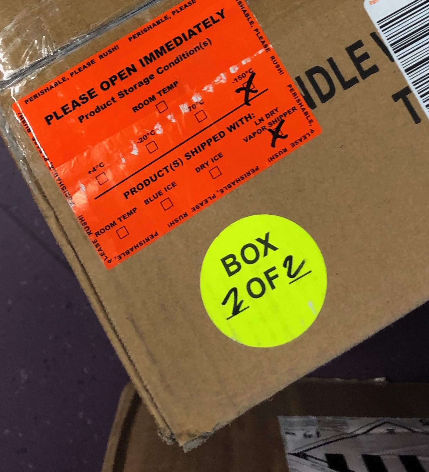
Please open immediately: shipped at minus 180 degrees
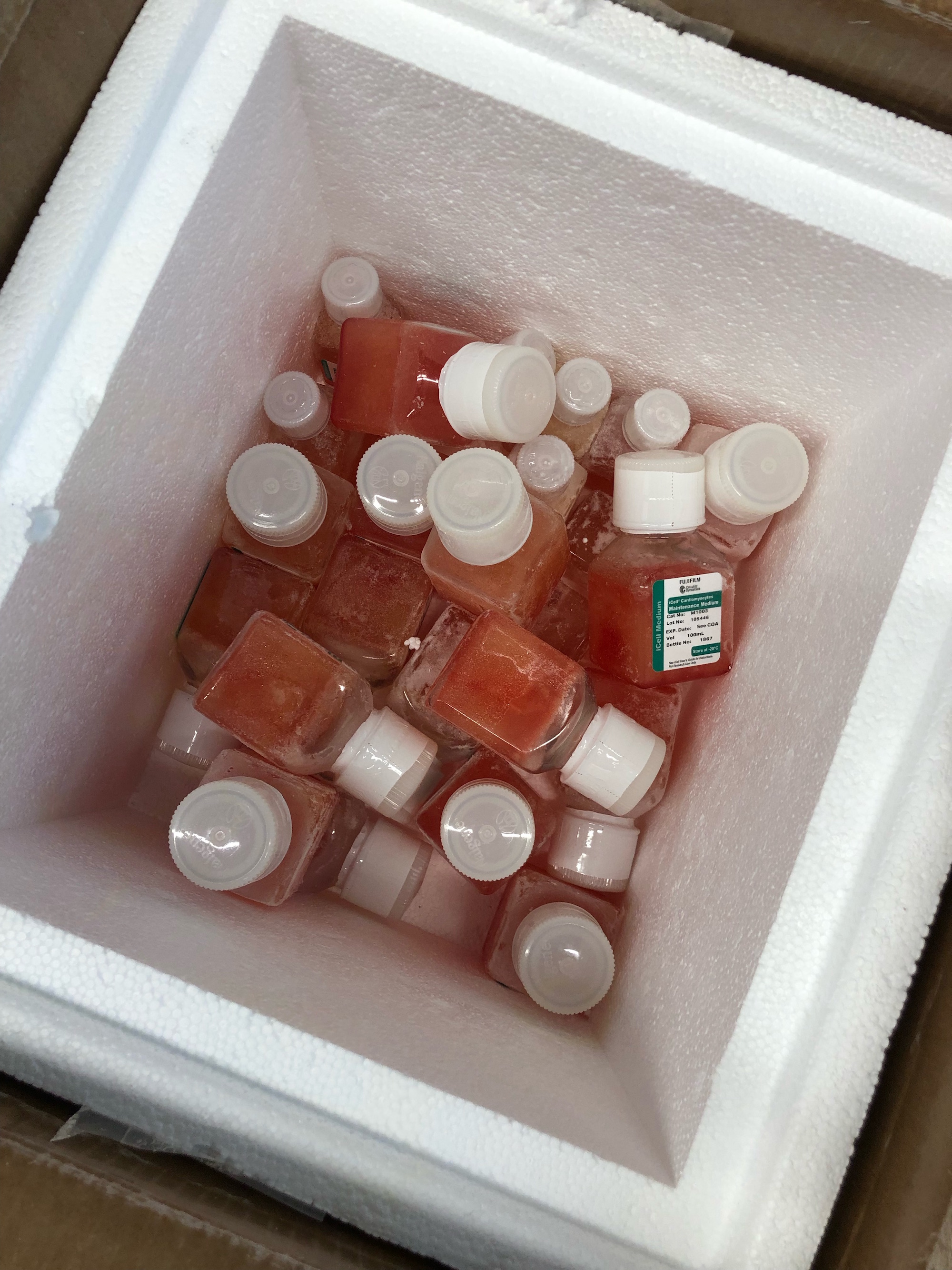
Bottles of media – just in time
Cell vials in protective canister [photos: Carmine Gentile]
Saturday, 11 September 2021
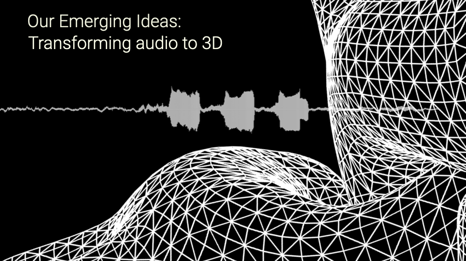
We have been working with the audio recording of George’s last breaths.
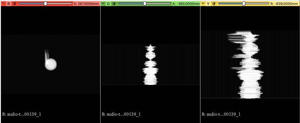
– translating the audio in different ways, with javascript, to generate animated imagery
– taking that animation as a sequence of still frames ( just as an MRI scan is a series of still frames) into 3D slicer
– using 3D Slicer to generate 3D form
– exporting STL files
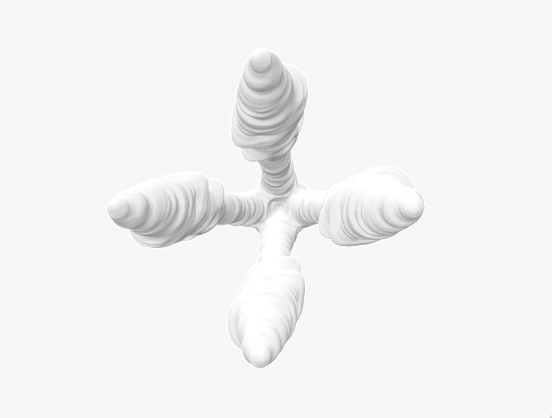
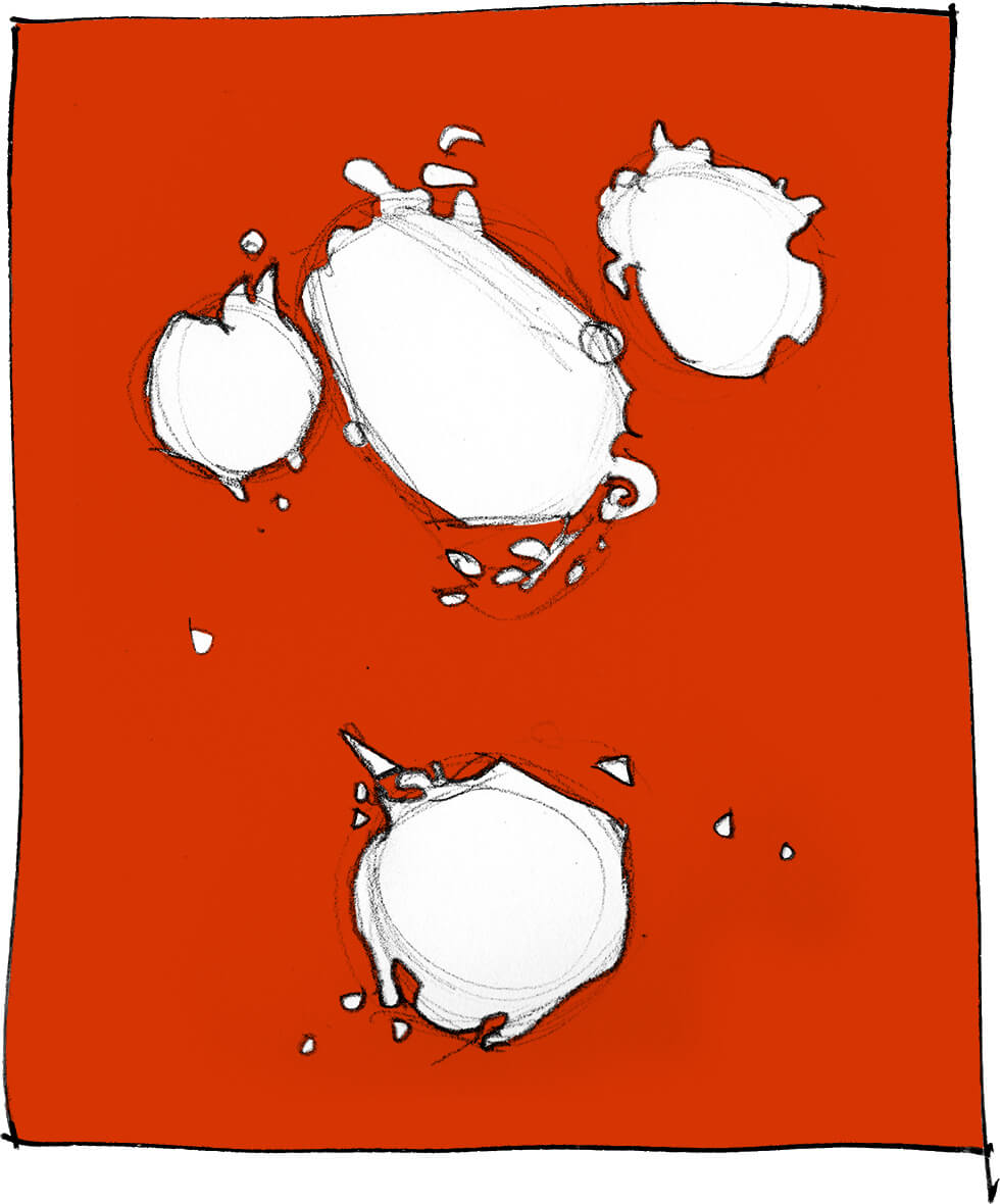
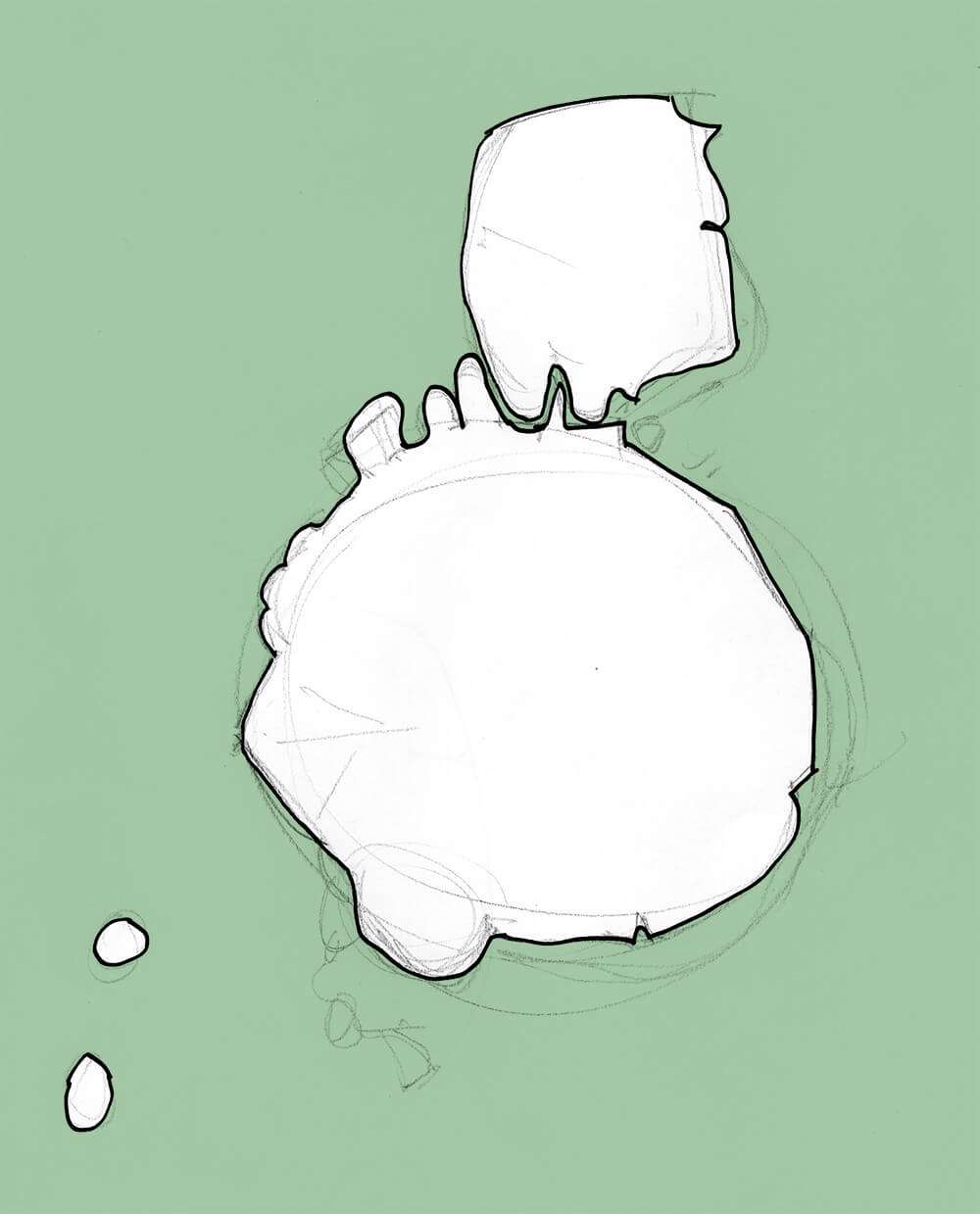
George’s breath as 3d shape, from frequency x volume over time
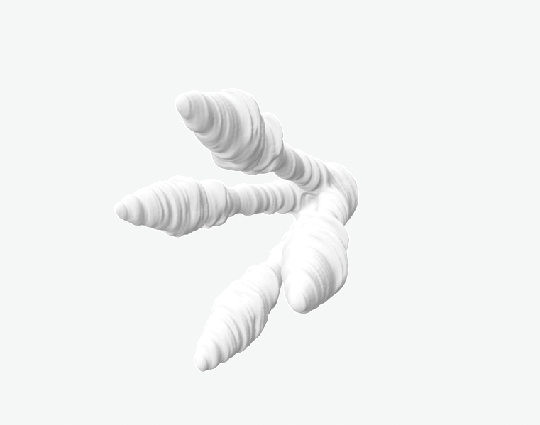
George’s breath as 3d shape, from frequency x volume over time
–> We are at the stage now of ordering millions of cells for future bio-printing of a form. We anticipate it will be tiny, a few cm high, given the number of cells we are working with.
–> We’re also interested in hearing and making audible, the cells’ beating
Saturday, 11 September 2021
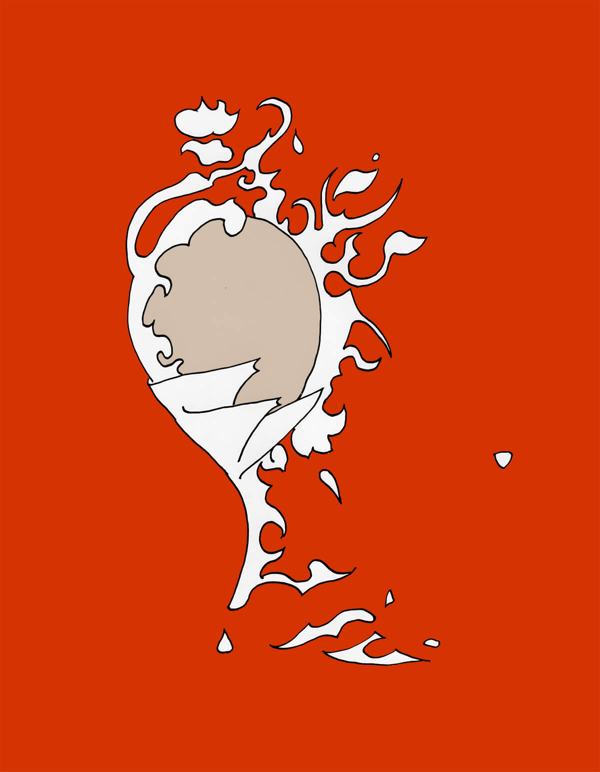
Linda’s friend and mentor, the artist George Schwarz, departed this life 28/5/2021. He had heart failure. Linda recorded some of his last breaths.
We are working with this recording to produce 3D form to be bio-printed with live, beating, cardiac spheroids – cardiac growth on the shape and structure of cardiac failure – new life forming from last breaths.
George is survived by his wife and co-conspirator, Charis, who is in close communication for this. See their work at http://www.george-charis.net
We have been through many different kinds of visualisations of the recording and printed some small tests on beautiful Hahnemuhle paper. In lockdown while most of life is online on screen, having a physical material surface seems called for.
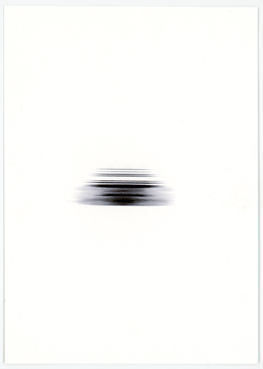
George’s breath, as lines
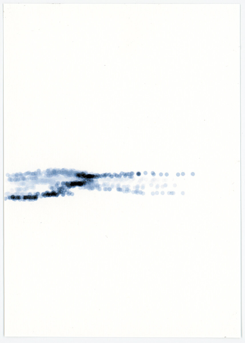
George’s breath, frequency as circles
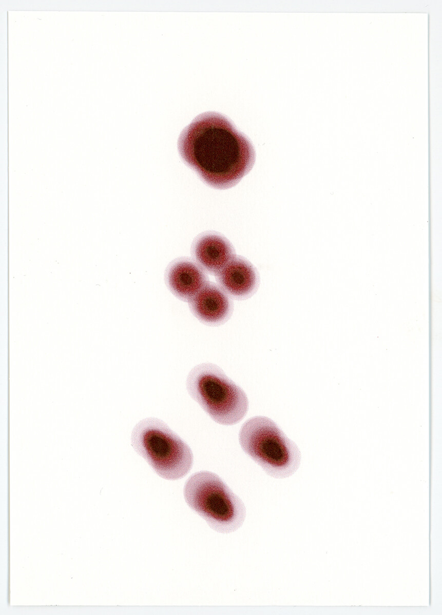
George’s breath, frequency x volume, at start, middle and end of duration
Friday, 27 August 2021
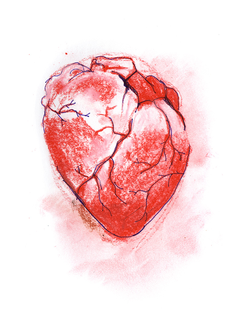
This week we have been much inspired by artist Melissa Coote’s heart paintings and bronze hearts.
“This arresting painting presents as its subject the most vital of all our organs, the heart. How strange and wonderful to behold the heart as if it were some kind of topographical landscape. We look down upon it as if it were an atoll isolated in the darkest sea. The undulations and ravines of its muscularity seem formed by the earth itself over thousands of years.”
from melissacoote.com
Thursday, 26 August 2021
Artist Horst Kiechle works with intricate accurate folded paper forms
He has also made a full paper folded torso with all organs, including the heart. Patterns are available online and have been downloaded and built by many.
https://www.instagram.com/explore/tags/papertorsobyothers/
Paper hearts by Horst Kiechle
Thursday, 19 August 2021
Carmine Gentile, our collaborator and project host, made a fantastic webinar presentation for Science Week on Monday, and Linda and I added a short presentation about our arts-science collaboration. Many thanks to Lynn Hutchinson and the crew at UTS Faculty of Science.
We had a great audience of around 350 attendees, and about 50 people put questions to Carmine, with some for us. Feedback was very positive. Here’s just one…
“Thank you very much for an excellent presentation and the opportunity to share in the fruits of the team’s painstaking research… The future of mankind is in good hands.”
For more on the Science Week presentation, and the recording of the presentation GO TO:
Mending Broken Hearts using Stem Cells and 3D Bioprinting Technology
Also available on Youtube here.
Sharing responses to two of the audience questions:
Linda: There are strong similarities in scientific research process and creative practice. Both address the unknown and often carry out material experiments to discover something new and to refine and clarify results. Creative practice can draw on widely eccentric sources in forming a work and so perhaps bring unexpected perspectives to the scientific which may spark other lines of enquiry or offer more layered ways of communicating, through aesthetics, feeling, multimedia, symbol, metaphor, storytelling and so on.
Linda: These are impossible to predict. For example, in our previous collaboration in 2020, by making a human life sized patch for the artwork prompted the researchers to try out patches at that scale, which they hadn’t before. Practical material issues of scale, flexibility and accurate fitting were investigated. Art practice can take unexpected approaches and so open up questions and avenues that might otherwise not be considered.
Saturday, 14 August 2021
Example of immaterial data being translated to 3D form – Artist Pierre Huyghe’s UUmwelt – cast from a 3D print of a visualisation of human thought as interpreted by neural net AI.
Pierre Huyghe: UUmwelt (Deep Image Reconstruction)
“… a set of elementary components, building blocks of ideas, were selected for a speculative situation. … They were given as images or descriptions to be imagined by a subject. As the person imagined these components, the brain activity was captured by an fMRI scanner and a deep neural network then attempted to reconstruct these thoughts, or ‘mental images’.”
This helps us think about the forms our audio might take once bio-printed.
Friday, 13 August 2021
We’ve been excitedly preparing for next Monday’s presentation for Science Week and Sydney Science Trail.
Our collaborator Carmine Gentile from the UTS Cardiovascular Regeneration Lab is leading a webinar titled “Science in Focus: Mending Broken Hearts with Cells and Bioprinting Technology”; and we are presenting an introduction to our project.
It’s coming right up… Monday 16, August, 2021, 12:30 – 1:30pm
And it’s not too late to register… Here
Sunday, 1 August 2021
We are still trying out ways of translating audio into sequences of frames of greyscale information, which can then be interpreted (like slices of MRI & CT scans are in 3D Slicer) into 3D form that could be bioprinted.
For the audio, we are staying close to the human experience of heart troubles – the sound patients hear when in the MRI scanner; the sound of human breath in a hospital ward; EEG signals from a person’s heart …
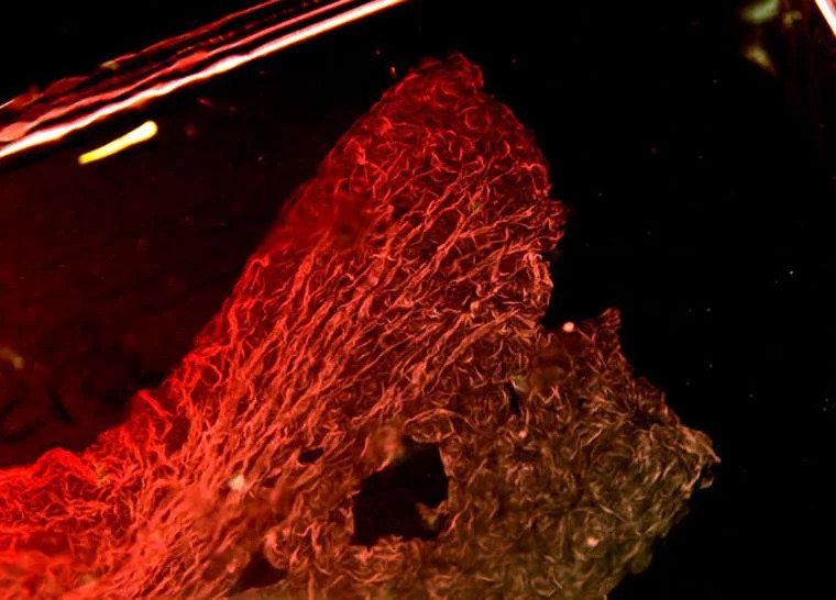
And more lockdown drawings of cardiac spheroids…
drawn from an image by Clara Liu Chung Ming: Mini-heart (healthy/control model), 2020
which are looking like they’d lend themselves nicely to tea towels, greeting cards and other merch, or is this the beginning of a graphic novel?
Friday, 23 July 2021
Inspired by the pared back, stylised woodblock prints in the Kaishi Hen I’ve been drawing cardiac spheroids.
drawn from 3D rendering analysis of a cardiac spheroid, that has an endothelial cell network that extends from the bioprinted cardiac spheroid into the hydrogel.
drawn from video recording of beating cardiac spheroids
Based on images in: https://iopscience.iop.org/article/10.1088/1758-5090/ac14ca/meta
Friday, 16 July 2021
Don’t hold us to this, but….
In cultivation of heart tissue three types of cells are needed :
These need to be in a certain ratio in the mix.
The cells communicate with each other, find each other, organise and join up and grow to make a spheroid.
We might be able to use this to inspire our creative methodology.
Thursday, 15 July 2021
We’ve continued our online meetings with Dr. Carmine Gentile and the team.
Today we were able to speak with a cardio thoracic surgeon for his perspective on this new research into cultivation and 3D printing of heart tissue. He showed us a Russian copy of History of the Heart – a book with imagery of the heart from Medieval times to the present. Glorious.
Each of the meetings has left us teetering between a kind of exuberant overwhelm & bright inspiration, also each meeting delivers a hundred related paths to follow.
We continue to read research papers, look at microscopy, at renderings and gather definitions for the millions of terms that are new to us. We have also been looking at some early work of anatomical imaging of the heart – Vesalius from 16th century Europe and Aoki Shukuya from 18th Century Japan.
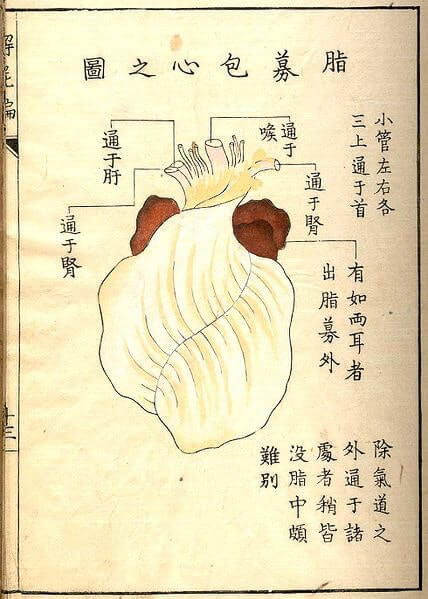
A woodcut by Aoki Shukuya from Kaishi Hen (Analysis of Cadavers), an anatomical atlas from the dawn of experimental medicine in Japan, published in Kyoto in 1772 – Source.
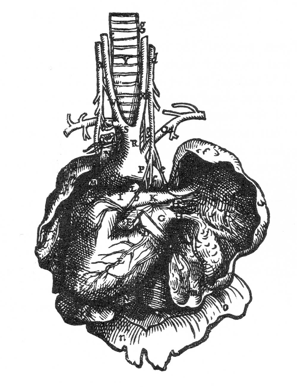
Heart by Andreas Vesalius from Dei Humani Corporus Fabrica 6th Book, 1543
Friday, 9 July 2021
Trying out the open source 3D Slicer software that Niina showed us. It’s for generating 3D models from MRI or CT scans. The software allows us to define areas from greyscale imagery to be turned into 3D forms. Today we’ve tried it out on non-medical images, experimenting with giving 3D form to other kinds of data– in this test: audio volume and frequency from voice recording.
It would be good to look at other data sources than this test audio visualisation.
— What data? What parallel or colliding or expanding fields / stories / processes could we use?
— What 3D form might be computationally generated / grown from our data?
Thursday, 1 July 2021
Our lab adventure has been cut short by lockdown.
We have changed tack & are having a series of one to one zoom meetings with each member of the team to find out more about their research projects. Each discussion so far has opened out whole worlds of fascinating detail and complexity:
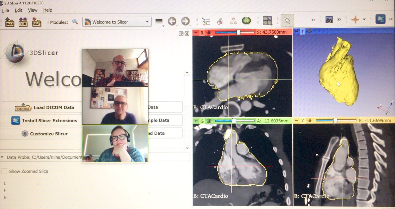
screenshot from our meeting with Niina Matthews who is working on 3D models from MRI and CT scans. These heart scans are public domain examples that come with the software.
Wednesday, 23 June 2021
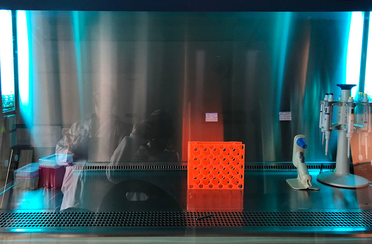
This morning we had an introduction to one of the labs– bio-printers, freezers, incubators etc with Dr. Carmine Gentile.
Friday, 18 June 2021
Artist Cynthia Verspaget, back in 2004 created the Anarchy Cell Line (TAnCL). This is a line of cells based on Cynthia’s own blood and HeLa cells. HeLa cells are commonly purchased for experimentation and used in labs, their originary cells were taken without consent from African American Henrietta Lacks in the 1950s.
“…the issues, concerns and questions I had concerning tissue ownership, lab techniques and the production of systems which set about disconnecting the social from the petri dish.
… Ambiguous boundaries were revealed through the very act of demarcation via the deliberate manipulation of cells, separated from the body. These categories found firstly instinctively and eventually through contemplation, are also shared by other symbolic monsters like the zombie (living – dead, human – non human).
HeLa had already been called monstrous, ‘combinations’ (and the less ambiguous term ‘hybrids’) are monstrous (HeLa and Cynthia is a combination creation), women’s bodies are leaky – monstrous, ‘othered’ bodies are monstrous… Rather than these terms being a rationale for expulsion, I found they offered potential for those that dwell in more than one category to break down boundaries not reinforce them. Monsters are filled with the potential for change, undoing, and opening up distinctions. Not only was there an ethical issue with the history of the procurement of HeLa cell line but there was an opportunity to seek out connections, to identify intersections for potential revelation and to perhaps seek out new metaphors to facilitate interrogation of these processes.”
Cynthia Verspaget
https://www.cynthiaverspaget.com/the-anarchy-cell-line
Wednesday, 16 June 2021
This week, we’ve been focused on research, and gathering ideas – our own and inspiration from others. For example….
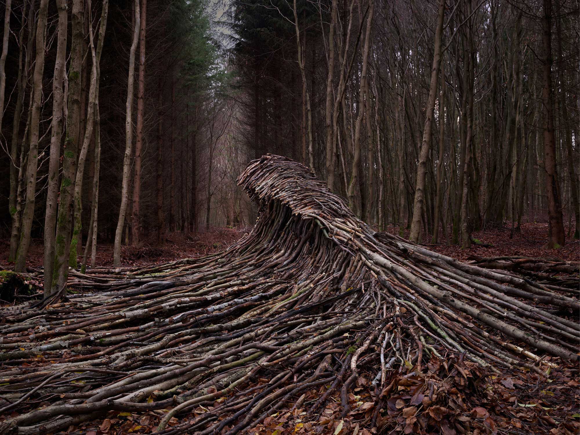
Jorg Glaescher, photographer, has been piling up sticks in the forest to produce forms.
— One of the aspects of our heart project is also the re assembly and creation of new form, from biological single units (cells in our case rather than sticks)
Image from Jorg Galschaer’s site: glaescher.de
Wednesday, 16 June 2021
Our research has also led us to…
Finding inspiration in the work of Neri Oxman of the Mediated Matter group at MIT who invent and fabricate material structures using biological processes – a silk pavillion spun by over 17,000 silk worms; an architectural surface with melanin that darkens in sunlight and clears in shade; programmable water based bio-composites; 3D printed glass; swarms of small 3D printing robots…. They also 3D print various materials in forms grown computationally from data.
— What forms might we grow for our heart project, and from what data?
TOTEMS – “3D printed to include six distinct liquid channels and pockets. Each pocket contains melanin from a different species, from bird to cuttlefish.”
from: The Mediated Matter Group https://www.media.mit.edu/projects/biodiversity/overview/
Tuesday, 8 June 2021
We’re meeting at UTS – a week for setting things up, ironing out admin and devising a schedule for the next 4 months
We’ve begun consultation with a couple of the researchers in the Cardiovascular Regeneration lab, tuning into the activities of the team. Their research for a paper is underway looking at the challenges of delivering 3D printed cardiac tissue. We have a flow chart to consider… the lab’s experiments in producing heart patches that will be viable during surgery.
We’ve commenced our related research. We need to get up to speed with the work of the lab… maybe we should frame this as an exploration of ‘Life amongst the Scientists’ (title of a well known book about laboratory life).
— How will our hybrid language of art & science form in this context of heart research?
— What does it look like as stem cells differentiate (transubstantiate) into heart cells?
— What happens throughout a heart attack in a petrie dish?
— When will our swipe cards arrive?
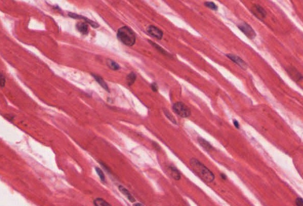
OpenStax College, CC BY 3.0 <https://creativecommons.org/licenses/by/3.0>, via Wikimedia Commons